PERIODONTAL POCKET OR GUM POCKET

THE PERIODONTAL POCKET

THE PERIODONTAL POCKET/DENTAL POCKET/GUM POCKETS/TEETH POCKET/POCKET FORMATION/TEETH GUM POCKETS/ PSUDOPOCKETS

WHAT DO PERIODONTAL POCKET LOOK LIKE..??

DEFINITION:

  • The Periodontal pocket is defined as a pathologically deepened gingiva sulcus.
  • A Periodontal Pocket is a fissure between the cementum and the epithelium and is created by the destruction of periodontal attachment apparatus.
  • Periodontal Pocket is roentgenolucent and not recorded on x-ray.

How to probe the periodontal pocket.??

Pockets are found by probing the gingival crevice around the entire circumference of the tooth.

Pocket depth can be recorded on the x-ray by inserting roentgenopaque material such as Gutta-Percha points into the pocket.

PERIODONTAL POCKET OR GUM POCKET

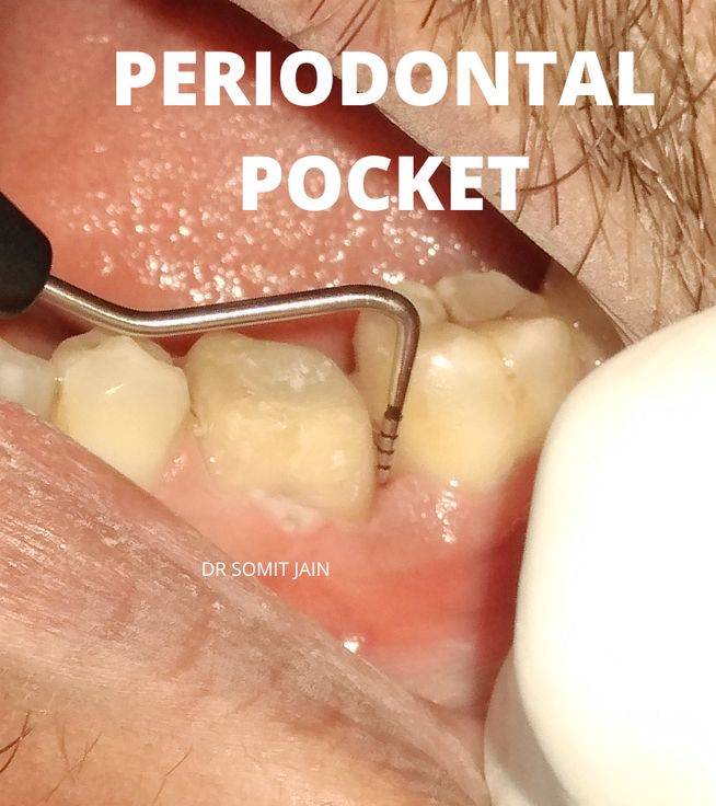
Illustration of Periodontal probing of a molar (Infrabony Pocket depth here recorded was 11 mm)

LIP-MUCOCELE-SWELLING-LASER

Treatment of Lower Lip Mucocele (lips swelling) with Diode laser

Treatment of Lower Lip Mucocele (lips swelling) with Diode laser Surgery

Mucoceles are common lesions of the oral mucous membrane involving minor salivary gland tissue. The lower lip is the site most frequently affected, the remainder occur in the cheek, palate, floor of the mouth, tongue, and retromolar fossa. The upper lip is rarely affected.
Mucoceles occur most often in children and young adults.

The mucocele, a mucus accumulation phenomenon of the salivary gland, is a common lesion of the oral cavity.

In the past it was believed that oral mucoceles arise from obstruction of an excretory duct, which caused back pressure of mucus and the formation of an epithelial-lined cyst. It is now, however, generally accepted that most mucoceles are caused by trauma-injury or severing of an excretory duct and subsequent escape of mucus into the adjacent tissue.

Histopathologically, there are two types of mucoceles:

1) Mucous extravasation cyst, generally regarded as being of traumatic origin, such as lip biting.

2) Mucous retention cyst result from obstruction of the duct of a minor or accessory gland.


Mucocele is a common lesion of the oral mucosa that results from an alteration of minor salivary glands due to a mucous accumulation. Mucocele involves mucin accumulation causing limited swelling

(1). Two types of mucocele can appear – extravasation and retention.

Extravasation mucocele results from a broken salivary glands duct and the consequent spillage into the soft tissues around this gland. Retention mucocele appears due to a decrease or absence of glandular secretion produced by blockage of the salivary gland ducts

(2). When located on the floor of the mouth these lesions are called ranulas because the inflammation resembles the cheeks of a frog

(3). Mucocele is a common lesion and affects the general population. For this reason we felt it would be interesting review the clinical characteristics of mucoceles, and their treatment and evolution in order to aid decision making in daily clinical practice.

LIP-MUCOCELE-SWELLING-LASER

MUCOCELE TREATMENT WITH DIODE LASER

MUCOCELE
ANKYLOGLOSSIA OR TONGUE TIE

Tongue-tie (Ankyloglossia): Symptoms, Causes, Diagnosis & Treatment

ANKYLOGLOSSIA OR TONGUE TIE

Lingual Frenectomy for Adults and baby with Diode Laser Surgical Procedure

Frenectomy of tongue

Tongue-tie which is also known as Ankyloglossia , is a congenital anomaly characterized by an abnormally short lingual frenulum which may restrict mobility of the tongue tip.

The phenotype varies from absence of clinical significance to rare complete ankyloglossia where the ventral part of the tongue is fused to the floor of the mouth.

The abnormally short lingual frenulum may result varying degree of decreased tongue mobility.

Tongue-tie (Ankyloglossia) has been suggested to cause breast-feeding difficulties (sore nipples, poor infant weight gain, early weaning), speech disorders (impaired articulation), problems with deglutition and dentition, oral-motor dysfunction and social issues related to the limited function of the tongue. (Lauren et al)

ANKYLOGLOSSIA OR TONGUE TIE

ANKYLOGLOSSIA OR TONGUE TIE

ANKYLOGLOSSIA OR TONGUE TIE

ANKYLOGLOSSIA-TONGUE TIE

Restricted tongue movement on protrusion

ANKYLOGLOSSIA OR TONGUE TIE RESTRICTED MOVEMENT

A LINGUAL FRENECTOMY (also known as a tongue-tie release) is the removal of a band of tissue (the lingual frenulum) connecting the underside of the tongue with the floor of the mouth.

A lingual frenectomy is performed to correct ankyloglossia (tongue-tie).

ANKYLOGLOSSIA OR TONGUE TIE surgery with Diode Laser

ANKYLOGLOSSIA OR TONGUE TIE DIODE LASER LINGUAL FRENECTOMY

ANKYLOGLOSSIA OR TONGUE TIE surgery with Diode Laser (Follow Up)

ANKYLOGLOSSIA OR TONGUE TIE SURGERY WITH DIODE LASER

Treatment of ANKYLOGLOSSIA OR TONGUE TIE with Diode Laser surgery (Infographic)

tongue-tie or Ankyloglossia Infographic
GINGIVAL DEPIGMENTATION DIODE LASER VS SCALPEL BLADE

Gingival depigmentation: Diode laser Vs Surgical blade; before and after.

GINGIVAL DEPIGMENTATION PROCEDURE (DIODE LASER VS SCALPEL BLADE) SMILE AMAZON DENTISTRY (मसूड़ों का कालापन कैसे दूर करे)

  • A smile expresses a feeling of joy,success, sensuality, affection and courtesy, and can reflect self confidence and kindness.
  • The harmony of a smile is not only determined by the shape, position, and color of the teeth, but also by the gingival tissues.
  • Gingival health and appearance are essential components of an attractive smile.
  • The color of the attached and marginal gingiva is generally described as coral pink. It is determined by several factors, including the number and size of blood vessels, epithelial thickness, quantity of keratinization and pigments within the epithelium.
  • Gingival melanin pigmentation (GMP) may occur as a result of several physiologic and/or pathologic factors, the most common of which is the normal physiologic production of melanin.
  • Gingival hyperpigmentation presents as a diffuse deep-purplish discoloration or brown and light-brown patches with irregular contours that do not alter the normal oral architecture.
  • GMP may result in complaints about poor esthetics, especially among individuals with excessive gingival display (GUMMY SMILE ).

Gingival depigmentation has been carried out using many procedures such as 

  • Scalpel surgery,
  • Gingivectomy,
  • Gingivectomy with free gingival autografting,
  • Cryosurgery,
  • Electrosurgery,
  • Chemical agents such as 90% phenol and 95% alcohol,
  • Abrasion with diamond burs,
  • Different types of LASERS
  • Different lasers include carbon dioxide (CO2) (10,600 nm), diode (820 nm), neodymium-doped:yttrium, aluminum, and garnet (Nd:YAG) (1,064 nm), erbium (Er)-doped:YAG (2,940 nm), and erbium- and chromium-doped:yttrium, scandium, gallium, garnet (Er,Cr:YSGG) (2,780 nm) lasers.

GINGIVAL DEPIGMENTATION PROCEDURE (DIODE LASER VS SCALPEL BLADE)

GINGIVAL DEPIGMENTATION DIODE LASER VS SCALPEL BLADE

GINGIVAL DEPIGMENTATION WITH DIODE LASER (SMILE AMAZON DENTISTRY)

Cosmetic Surgery in Raipur

SMILE AMAZON DENTISTRY GINGIVAL DEPIGMENTATION

GUM BLEACHING

GUM BLEACHING

GINGIVAL DEPIGMENTATION: SPLIT MOUTH
1. RIGHT: SURGICAL BLADE 2. LEFT: DIODE LASER

GINGIVAL DEPIGMENTATION- SPLIT MOUTH

GINGIVAL DEPIGMENTATION WITH LASER

GINGIVAL DEPIGMENTATION WITH LASER

GINGIVAL DEPIGMENTATION VIDEO SHOWING BEFORE AND AFTER

DENTAL PLAQUE-TARTAR-CALCULUS

Is DENTAL PLAQUE a BIOFILM..??

Is DENTAL PLAQUE a Biofilm..??

Dental Plaque

WHO definition of Dental Plaque:

  • Dental plaque has been defined as “a specific but highly variable structural entity consisting of micro-organisms and their products on tooth surfaces, restorations and other parts of oral cavity composed of salivary components like mucin, desquamated epithelial cells, debris and micro-organisms, all embedded in a highly organized extracellular matrix.” (WHO-1961)
  • Dental plaque biofilm is a complex, highly organized three dimensional community arrangements of microorganisms involved in a wide variety of physical metabolic and molecular interaction that adhere to surfaces where there are moisture and nutrients.
  • Dental plaque represents a true Biofilm consisting of a variety of micro-organisms involved in a wide range of physical, metabolic and molecular interactions.
  • Plaques are usually colorless sticky bacterial film and hence DISCLOSING SOLUTIONS have become popular to enhance visualization. A red stained mat on the labial or buccal surface of a tooth is the prevalent image of the bacterial plaque.
  • The bacterial plaque biofilm is the pathogenic locus of both DENTAL CARIES and inflammatory PERIODONTAL/GUM disease.

Picture displaying: Dental plaque build up on teeth and gumline area

DENTAL PLAQUE-TARTAR-CALCULUS

DENTAL PLAQUE-TARTAR-CALCULUS

NATURE OF DENTAL PLAQUE

  • Using the new tools available to us we find that the plaque biofilm is not a polyglot mass but rather specific types of MICROCOLONIES surrounded by a gel-like intercellular substance derived from the bacteria themselves, from saliva, from the gingival crevicular fluid (GCF) and the inflammatory exudate.
  • The matrix serves as the pathway for fluid movement within the plaque and contains the products of plaque metabolism (acids,enzymes,toxins etc); salivary constituents (proteins, carbohydrates and salts); soluble food components; shed leukocytes and epithelial cells.

DENTAL PLAQUE FORMATION : DYNAMICS

Plaque is not an accidental accumulation of bacteria. It forms in a sequence of steps

STEP 1: PELLICLE FORMATION

  • Deposition of PELLICLE, a cuticular like material derived Supragingivally from saliva and Subgingivally from gingival crevicular fluid (GCF).
  • Salivary pellicle is composed mainly of glycoproteins (a mucoid type of protein containing carbohydrate) including a high molecular weight material called the “Agglutinating substance”
  • This pellicle begins to form immediately after brushing, is very tenacious and can attract and help anchor specific kinds of bacteria to the tooth surface.
STEP 2: BACTERIAL COLONIZATION
 
  • The attachement of specific species of Gram positive Streptococci to the pellicle surface is called bacterial colonization.
  • These early plaques can be highly cariogenic.
  • The bacteria grow divide and produce an extracellular gel called DEXTRAN, which help anchor the growing mass to the tooth and attracts and traps other bacterial forms.
  • Within a few days a mixture of different types of microorganism comprise the bacterial community.
  • As the planktonic microorganisms multiply and grow, they form stratified layers against the tooth surface. Filamentous microorganisms grow on the surfaces of the coccal colonies and begin to replace the cocci.
STEP 3: PLAQUE MATURATION (Biofilm development and growth)
 
  • The development of complex flora-plaque maturation is the next step in the formation of dental plaque biofilm.
  • The microorganisms in the community will secret extracellular polysaccharides which are produced by the bacteria in response to sucrose and fermentable carbohydrates.
  • this mature plaque is responsible for the initiation of Periodontal/Gum disease.

FORMATION OF CALCULUS:

  • In areas opposite the salivary glands and in subgingival areas where the fluids are rich in calcium and phosphates, the deeper layers of bacteria can become mineralized.
  • Individual foci of calcification form, enlarge and coalesce and calculus forms.
  • The mineralized layers of plaque are always covered by unmineralized plaque so that the formation of calculus is a special aspect of plaque formation and not a separate process.